H. pylori Antigens Rapid Test Device Sample Feces Rapid Test Strip Kit
INTENDED USE
The H. pylori Antigen Rapid Test Device (Feces) is a rapid visual immunoassay for the qualitative presumptive detection of Helicobacter pylori antigens in human fecal specimens. This kit is intended to be used as an aid in the diagnosis of H. pylori infection.
INTRODUCTION
Helicobacter pylori (also known as Campylobacter pylori) is a spiral-shaped with a typical flagellum, Gram negative bacteria, infecting gastric mucosa. It causes several gastro-enteric diseases such as non-ulcerous dyspepsia, gastric and duodenal ulcer, active gastritis and can even increase the risk of stomach adenocarcinoma, so as to be classified as carcinogen agent type I.
Many H. pylori strains have been isolated: among them, the strain expressing CagA antigen is strongly immunogenic and, according to this, it is of utmost clinical importance because it is associated to the cytotoxic factor. It is widely reported in many literature articles that, in infected patients showing antibodies against CagA gene product, the risk of gastric cancer is up to five times higher than the reference group infected with a CagA negative bacterial strain.
The presence of the gene itself determines the persistence of the infection, the ulceration and the protein associated, VacA toxin is frequently the main cause of infiltrations in the gastric mucosa.
This antigen associated to others, such as CagII, CagC, seems to act as starting agent of a sudden inflammatory response which can provoke ulceration (peptic ulcer), allergic episodes, and a decrease of the therapy efficacy.
At present several invasive and non-invasive approaches are available to detect this infection state.
Invasive methodologies requires endoscopy of the gastric mucosa with a histologic, cultural and urease investigation, which are cost-effective and requiring long times to come to a correct final diagnosis.
Alternatively, non-invasive methods are available such as Breath Test, which is extremely complicated and not highly selective, or classical ELISA and immunoblotting assays.
MAIN CONTENTS
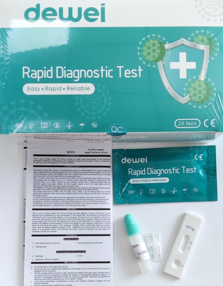
• One pouch containing a reaction test Cassette with desiccant.
• Specimens dilution tube with buffer.
• Instructions for use.
STORAGE AND STABILITY
• Store at 39~ 86 º F (4 ~ 30 º C) in the sealed pouch for 18 months.
PRECAUTIONS
• For in vitro diagnostic use only.
• Do not use after expiration date.
• The test Cassette should remain in the sealed pouch until use.
• The used test Cassette should be discarded according to local regulations.
DIRECTION OF USE
Bring tests, specimens, buffer and/or controls to room temperature (15-30°C) before use.
Specimen collection and pre-treatment:
Best results will be obtained if the assay is performed within 6 hours after collection.
Unscrew and remove the dilution tube applicator. Be careful not to spill or spatter solution from the tube. Collect specimens by inserting the applicator stick into at least 3 different sites of the feces to collect approximately 50 mg of feces (equivalent to 1/4 of a pea).
Place the applicator back into the tube and screw the cap tightly. Be careful not to break the tip of the dilution tube.
Shake the specimen collection tube vigorously to mix the specimen and the extraction buffer.
Testing:
Remove the test from its sealed pouch, and place it on a clean, level surface. Label the test with patient or control identification. To obtain a best result, the assay should be performed within one hour.
Using a piece of tissue paper, break the tip of the dilution tube. Hold the tube vertically and dispense 2 drops of solution into the specimen well (S) of the test device.
Avoid trapping air bubbles in the specimen well (S), and do not drop any solution in observation window.
As the test begins to work, you will see color move across the membrane.
Wait for the colored band(s) to appear. The result should be read at 10 minutes. Do not interpret the result after 20 minutes.
Note:
If the specimen does not migrate (presence of particles), centrifuge the extracted specimens contained in the extraction buffer vial. Collect 80 µL of supernatant, dispense into the specimen well (S) of a new test device and start afresh following the instructions mentioned above.
INTERPRETATION
POSITIVE: Two colored bands appear on the membrane. One band appears in the control region (C) and another band appears in the test region (T).
NEGATIVE: Only one colored band appears, in the control region (C). No apparent colored band appears in the test region (T).
INVALID: Control band fails to appear. Results from any test which has not produced a control band at the specified read time must be discarded. Please review the procedure and repeat with a new test. If the problem persists, discontinue using the kit immediately and contact your local distributor.
FRQ:
| 1 |
What is the cause of H pylori infection? |
H. pylori bacteria are usually passed from person to person through direct contact with saliva, vomit or stool. H. pylori may also be spread through contaminated food or water. The exact way H. pylori bacteria causes gastritis or a peptic ulcer in some people is still unknown. |
| 2 |
What are the signs and symptoms of H. pylori? |
H. pylori is a bacteria that can cause peptic ulcer disease and gastritis. It mostly occurs in children. Only 20% of those infected have symptoms. Symptoms include dull or burning stomach pain, unplanned weight loss and bloody vomit. |
| 3 |
Can H. pylori be cured completely? |
In some cases, H pylori can't be cured with any therapy, though the symptoms may be able to be reduced. If cured, reinfection may occur in areas where sanitary conditions are poor. |
| 4 |
How serious is H pylori infection? |
It can damage the tissue in your stomach and the first part of your small intestine (the duodenum). This can cause pain and inflammation. In some cases, it can also cause painful sores called peptic ulcers in your upper digestive tract. H. pylori is common. |
| 5 |
What do bowel movements look like with H. pylori? |
pylori infection, you might have blood in your stool. In most cases, the blood appears very dark — almost black. Your stools might have a tarry appearance or consistency. If you have bloody stools, you should call our office right away so we can get any bleeding under control. |
| 6 |
What not to eat when you have H. pylori? |
Citrus fruits such as lemons and pineapple can increase stomach acid and cause pain and heartburn. Spicy foods. Spicy or acidic foods like garlic, mustard and pepper can all have a negative effect on people with H. |
| 7 |
What is the fastest way to cure H. pylori? |
Ulcers caused by H. pylori are usually treated with a combination of antibiotics and a proton pump inhibitor (PPI). Triple therapy: Therapies that combine PPIs with two antibiotics remain the first-line options for treating H. pylori. |
| 8 |
What is the first stage of H. pylori? |
Although a staging system for the H pylori infection does not exist, some steps in the disease process are well described. The first step is chronic gastritis, followed after a time by the second step, atrophic gastritis. The third step is intestinal metaplasia, which may evolve into dysplasia. |
| 9 |
What happens if you test positive for H. pylori? |
A positive test result means that you have an H. pylori infection. Your provider will usually prescribe one or more antibiotics to treat the infection. You will usually take other medicines to relieve your symptoms and help heal your stomach. |
| 10 |
How contagious is H. pylori? |
H. pylori is commonly transmitted person-to-person by saliva. The bacteria can also be spread by fecal contamination of food or water. In developing countries, a combination of untreated water, crowded conditions, and poor hygiene contributes to higher H. |
| 11 |
Can your body get rid of H. pylori by itself? |
The almost invincible human immune system fails to eliminate H. pylori because of successful immune evasion strategies and also because of the complex intrinsic genetic variability adopted by this bacterium. |
| 12 |
Which probiotic kills H. pylori? |
pylori treatment reveals that single-strained probiotics, particularly those containing Lactobacillus spp., Bifidobacterium spp., and Saccharomyces spp. are more commonly used. These probiotics have shown promise in increasing the success rate of H. pylori eradication and in mitigating side effects. |
| 13 |
Can stress cause H. pylori? |
Certain health and lifestyle factors increase the risk of damage to the stomach and intestinal lining. These factors make it more likely that a person will develop an ulcer, including a stress-related ulcer: H. pylori infection. |
| 14 |
What is the natural killer of H. pylori? |
In human studies , daily consumption of broccoli sprouts reduced H. pylori colonization and gastric inflammation, with some cases resulting in complete eradication of the bacteria. |


 Your message must be between 20-3,000 characters!