Lymphatic Filariasis Antibody IgM/IgG Rapid Test Blood
INTENDED USE
The Filariasis IgG/IgM Rapid Test is a lateral flow immunoassay for the simultaneous detection and differentiation of IgG and IgM anti-lymphatic filarial parasites (Wuchereria Bancrofti, Brugia Malayi and Brugia Timori) in human serum, plasma or whole blood. This test is intended to be used as a screening test and as an aid in the diagnosis of infection with lymphatic filarial parasites. Any reactive specimen with the Filariasis IgG/IgM Rapid Test must be confirmed with alternative testing method(s).
For professional use only.
For in vitro diagnostic use only.
| Storage |
2~30 º C |
| Specimen |
Whole Blood/Serum/Plasma |
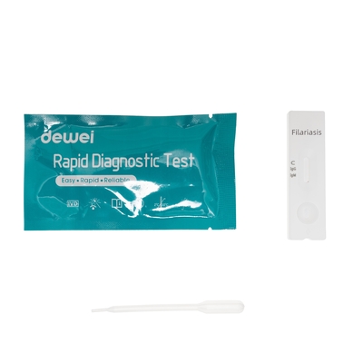
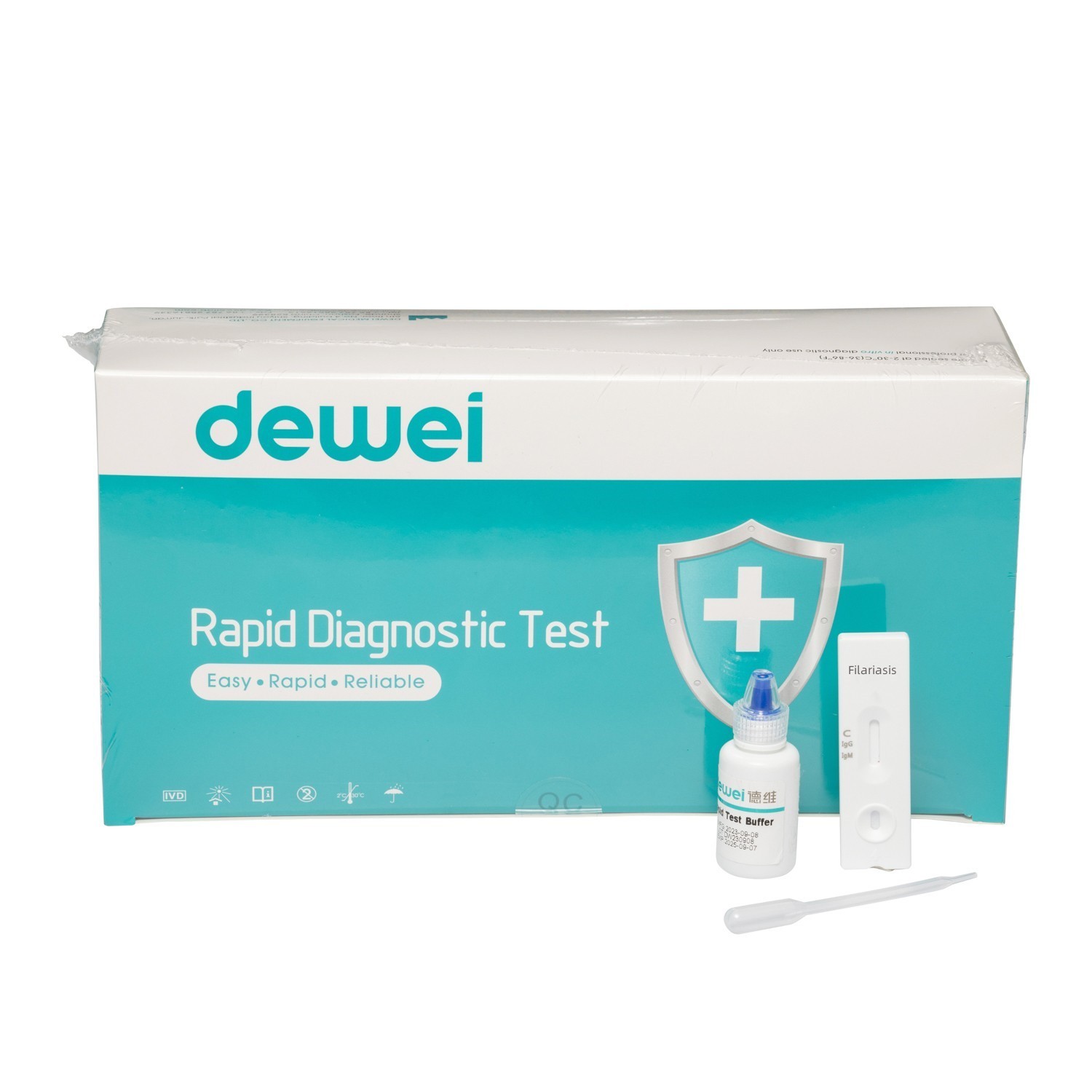
| Component |
Rapid Test +Buffer + Pipettes |
| Principle |
Colloidal gold rapid tests |
| Reading |
Within 15mins |
| Package |
40tests/box; 25tests/box |
| Trademark |
Dewei |
| Origin |
China |
INTRODUCTION
The lymphatic filariasis known as Elephantiasis, affects about 120 million people over 80 countries.
There are 3 types of these thread-like filarial worms:
Wuchereria bancrofti,
Brugia malayi,
Brugia timori.
MAIN CONTENTS
Rapid Test Cassette in Pouch
Buffer
Disposable dropper
Package insert
SRORAGE
Store as packaged in the sealed pouch at room temperature or refrigerated (2-30°C).
The test is stable through the expiration date printed on the sealed pouch.
The test must remain in the sealed pouch until use.
DO NOT FREEZE.
Do not use beyond the expiration date.
PRECAUTIONS
• For professional in vitro diagnostic use only.
• Do not use after the expiration date indicated on the package. Do not use the test if the foil pouch is damaged. Do not reuse tests.
• This kit contains products of animal origin. Certified knowledge of the origin and/or sanitary state of the animals does not completely guarantee the absence of transmissible pathogenic agents. It is therefore, recommended that these products be treated as potentially infectious, and handled by observing usual safety precautions (e.g., do not ingest or inhale).
• Avoid cross-contamination of specimens by using a new specimen collection container for each specimen obtained.
• Read the entire procedure carefully prior to testing.
• Do not eat, drink or smoke in the area where the specimens and kits are handled. Handle all specimens as if they contain infectious agents. Observe established precautions against microbiological hazards throughout the procedure and follow standard procedures for the proper disposal of specimens. Wear protective clothing such as laboratory coats, disposable gloves and eye protection when specimens are assayed.
• Humidity and temperature can adversely affect results.
• Used testing materials should be discarded according to local regulations.
OPERATION PROCEDURE
Bring tests, specimens, buffer and/or controls to room temperature (15-30°C) before use.
1) Remove the test from its sealed pouch, and place it on a clean, level surface. Label the device with patient or control identification. For best results the assay should be performed within one hour.
2) Testing
Transfer 1 drop of whole blood, serum or plasma sample to the specimen well (S) with the provided disposable pipette.
Then add 1 drop of buffer and start the timer.
Avoid trapping air bubbles in the specimen well (S), and do not add any solution to the result area.
As the test begins to work, color will migrate across the result area in the center of the device.
3) Wait for the colored band(s) to appear. The result should be read within 15 minutes. Do not interpret the result after 20 minutes.
FRQ:
1) What causes lymphatic filariasis?
Lymphatic filariasis is caused by infection with parasites classified as nematodes (roundworms) of the family Filariodidea. There are 3 types of these thread-like filarial worms: Wuchereria bancrofti, which is responsible for 90% of the cases. Brugia malayi, which causes most of the remainder of the cases.
2) Does lymphatic filariasis go away?
Is there a cure for lymphatic filariasis? There's no vaccine or cure for filariasis. Medication can kill many of the worms and keep you from spreading the infection to someone else. Treatment can also reduce filariasis symptoms.
3) What are the signs of filariasis?
Signs and symptoms:
Fever.
Inguinal or axillary lymphadenopathy.
Testicular and/or inguinal pain.
Skin exfoliation.
Limb or genital swelling - Repeated episodes of inflammation and lymphedema lead to lymphatic damage, chronic swelling, and elephantiasis of the legs, arms, scrotum, vulva, and breasts.
4) Which mosquito causes lymphatic filariasis?
Lymphatic filariasis is transmitted through the bite of infected Culex quinquefasciatus mosquito in Tamil Nadu. Brugia malayi is transmitted by Mansonia annulifera, M. uniformis and M. Indiana in Kerala.
5) How do I get rid of filarial worms?
Treatment overview:
The usual treatment for lymphatic filariasis is a drug called diethylcarbamazine (DEC), which kills both immature and adult parasitic worms. Your healthcare provider may recommend either a single-day treatment or taking the drug for 12 days. DEC has been used globally for more than 50 years.
6) Who is most likely to get lymphatic filariasis?
Lymphatic filariasis affects over 120 million people in 72 countries throughout the tropics and sub-tropics of Asia, Africa, the Western Pacific, and parts of the Caribbean and South America. Whereas the disease was once thought to affect only adults, it now appears that most infections are acquired in childhood.
7) How does filariasis start?
Filariasis is a disease caused by a chronic mosquito-borne parasitic infection. Chronic infection can lead to swelling of the extremities, hydroceles, and testicular masses. It is the second-largest cause of permanent deformity and disability behind leprosy worldwide.
For details, please contact Dewei staff for Instruction Manual!


 Your message must be between 20-3,000 characters!